


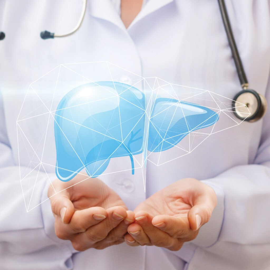
Liver Cancer
The liver is an organ located behind the abdominal cavity at the level of the lumbar vertebrae L1-L2, surrounded by various organs such as the stomach, kidneys, muscles, and small intestines. This causes tumors to often expand towards the back and spread to blood vessels and neighboring organs. The spread to important blood vessels and nearby organs indicates the severity and stage of the disease.
Diagnosis
Diagnosis Early diagnosis helps improve the patient's survival rate, but it is challenging to diagnose HCC in the early stages. Patients in the early stages often have vague symptoms, and primary care physicians may not consider it. Therefore, important and necessary diagnostic methods are computed tomography (CT) scans or magnetic resonance imaging (MRI). However, small lesions may not be detected in some cases, and there are limitations in patients with impaired kidney function or the inability to extract tissue for examination during the same period.
Currently, there are new non-invasive diagnostic methods that do not require surgery. The procedure is not complicated and similar to endoscopic examination of the stomach. This is called endoscopic ultrasonography (EUS), which has a small ultrasound device attached to the endoscope. This allows for better visualization of organs in the abdominal cavity, including the liver and bile ducts, which are located behind the stomach and initial portion of the small intestines. The images are usually clearer than conventional abdominal ultrasound examinations. Biopsy of the liver can be performed through the endoscope using a small needle, confirming the diagnosis at the same time. This method does not require contrast agents injected into the bloodstream, such as with CT scans or MRIs, which helps avoid adverse effects on kidney function.
Preparation before the Examination
1. Avoid all food and drinks for 6 hours before the examination.
2. Patients with chronic diseases can take medications according to the physician's treatment plan, except for diabetes medications (fasting on the day of the examination) and blood thinners (as instructed by the physician before the endoscopy).
3. In the case of endoscopy with biopsy, it may be necessary to stay in the hospital for at least 1 day for observation.
4. Please bring a companion.
5. At least 1 day before the appointment, the nurse will provide instructions and confirm the appointment by phone.
During the Examination
1. Vital signs will be assessed, and weight and height will be measured.
2. Change into a gown and remove dentures (if applicable).
3. Blood sampling or saline infusion may be required, depending on the physician's treatment plan.
4. The attending physician will provide instructions and evaluate readiness before entering the endoscopy room.
5. The endoscopy procedure takes approximately 30-60 minutes.
Post-examination Care
1. After the endoscopy, you will receive approximately 1 hour of recovery and observation in a resting room for your safety.
2. The physician will inform you and your family about the preliminary examination results. The results of the biopsy will take approximately 3 working days.
3. The nurse will assess readiness for transfer to the patient ward or discharge home according to safety criteria before leaving the endoscopy unit.
Things the patient should know or do after the examination
1. Mild throat discomfort may be felt on the first day, which can be relieved with lozenges.
2. There may be minimal bleeding at the site where the tissue biopsy was taken, but it does not have any significant impact on the patient.
3. Before going home, you will receive instructions on monitoring for abnormal symptoms and follow-up appointments, as well as a post-procedure phone call.
For additional information, please contact the Endoscopic Ultrasound Center at 1645, press 1, then press 2372.
Date 24.02.2023